|
|
|
|
Descriptions |
Genera |
|
 |
Apicomplexans
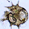
(sporozoa, telosporidea) Campbell p.555 They are single-cells symbiotrophs modified to penetrate tissue and obtain food from animals. All apicomplexans are parasites of animals and cause diseases like malaria. They can reproduce sexually and asexually, but these life cycles often require two or more host species for completion. |
Babesia Haplosporidium Toxoplasma |
 |
Brown
Algae (phaeophytes) Campbell p.560 They are the largest, complex protoctists with as much as 60 meters long. They dominate the intertidal zone, where they form great seaweed beds. The phaeophytes are crucial primary producers, providing habitat and food for other protoctists, marine animals, and microbes. |
Alaria Laminaria Seirococcus |
 |
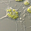
Cellular
Slime Molds Campbell p.565 The feeding stage consists of individual cells, but once food sources are depleted, many cells aggregate to function as a unit. They may seem similar to plasmodial slime mold, but each cells are distinct from one another by their membranes. Other differences include asexual reproduction, no flagellated stages, and being a haploid organism. |
Dictyostelium Mayorella Thecamoeba |
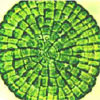 |
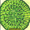
Charophycean
(green algae) Campbell p.567, 569, 573-74 This is the other group of green algae (chlorophytes), they are the closest relatives of land plants. Four key traits that correlate charophyceans with land plants are the rose-shaped complexes for cellulose synthesis, peroxisome enzymes, structure of flagellated sperm, and formation of phragmoplast. |
Chara Coleochaete |
 |
Chlorarachniophytes
Campbell p. 552 An alga that can be found in the tropical oceans. They are similar to the golden algae in that they are also mixotrophic, capable of feeding on bacteria and protists in the absence of light. They have cytoplasmic extensions to capture a prey by forming a net. |
|
 |
Chlorophytes
(green algae) Campbell p.567 They are algae that have grass-green chloroplasts surrounded by two membranes and that form zoospores or gametes having undulipodia, usually at least two, of equal length. They are a major component of the freshwater photoplankton; it has been estimated that they fix more than a billion tons of carbon every year. |
Chlamydomonas Pseudobryopsis Trebouxia |
 |
Choanoflagellate
(collar flagellates) Campbell p.628 Each choanoflagellate consists of one flagellum with hairlike rings surrounding the base. Most are sessile with the stalk connected to a surface. They resemble the collar cells of sponges, therefore, choanoflagellate may have given rise to the first true animal. |
Desmarella Kentrosiga Monosiga |
 |
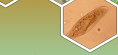
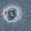
Ciliates
Campbell p.556 Most ciliates, which are among the best-known protoctists, are bactivorous (bacteria eaters) single cells. They possess two different types of nuclei, small micronuclei and larger macronuclei, usually more than one of each kind. Nearly all are phagotrophic, eating bacteria, tissue, or other protists, or they are osmotrophs, utilizing water nutrients. |
Didinium Prorodon Tetrahymena |
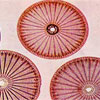 |
Diatoms
(Bacillariophyta) Campbell p.559 Each valve of the diatom test (shell) is composed of pectic organic materials impregnated with silica, in an opaline state. They can reduce the concentration of silica in the water below a level detectable by chemical techniques. They are one of the most important organisms at the base of marine and freshwater food chains. |
Achnanthes Fragilaria Tabellaria |
 |
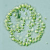
Dinoflagellates
Campbell p.555 Abundant in warm seas and most swim as members of marine plankton. Many species are reinforced by internal plates of cellulose. They move through the water by spinning caused by the two flagella. The phenomenon called "red tide" is caused by a massive population of dinoflagellates. |
Amphidinium Noctiluca Prorocentrum |
 |
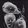
Diplomonads
Campbell p. 552 Diplomonads are two-celled organisms. They have two equal-sized nuclei and four flagella on each cell. They are parasitic flagellates, the most notorious is Giardia intestinalis, a parasite that inhabits the intestine of mammals ingested through contaminated water. |
Giardia Hexamita Trepomonas |
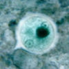 |
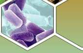
Entamoebas
Campbell p. 564 They are parasites that infect all vertebrates and some invertebrates. The cells have a single nucleus, a slightly protruding pseudopodia, and measuring at 10 to 100 µm in diameter. The most infamous genus, Entamoeba histolytica, causes amoebic dysentery in humans. |
Entamoeba Endolimax |
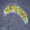 |
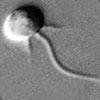
Euglenids
Campbell p.554 Euglenids have two distinct features: a pocket at one end where one or two flagella emerge, and a storage molecule called paramylon granule. Instead of a rigid cellulosic wall, they have pellicles covering the outside made up of protein. |
Euglena Hyalophacus Rhabdomonas |
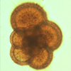 |
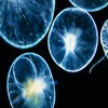
Foraminiferans
(forams) Campbell p.563 These organisms bear reticulopods, cells that fuse to form networks in which bidirectional streaming can be seen. Foraminiferans have pore-studded shells, or tests, hardened with calcium carbonate. Found in both the ocean and fresh water, but most live in the sand or attach themselves to rocks or algae. |
Allogromia Iridia Rotaliella |
 |
Golden
Algae (chrysophytes) Campbell p.560 |
Dinobryon Nematochrysis Trentonia |
 |
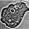
Gymnamoebas
Campbell p.564 They are free-living, shapless, unicellular organisms that feed by engulfing their prey (endocytosis). These unicellular protists are ubiquitous in soil as well as freshwater and marine environments. |
Acrasia Hyalodiscus Pocheina |
 |
Kinetoplastids
Campbell p.553 This group of organisms has a special large mitochondrion called the kinetoplast. These protists can be found in freshwater, marine, and moist terrestrial ecosystems. The inframous genus, Trypanosoma, causes sleeping sickness and Chagas' disease. |
Crithidia Naegleria Trypanosoma |
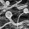 |
Oomycetes
(water molds, white rusts, downy mildews) Campbell p.558 Traditionally been considered fungi, since oomycete means "egg fungus." Their cell walls are made of cellulose, whereas chitin makes up the cell walls of fungus. They are either saprobes or symbiotrophs, they feed by extending funguslike threads, or hyphae, into victimized tissues, where they release digestive enzymes and absorb the resulting nutrients. |
|
 |
Parabasalids
Campbell p. 553 They are a group of flagellate protozoa dwelling symbiotically in animals. They are anaerobic, lacking mitochondria, and have a flagellum at their anterior ends. An infamous example of parabasalid is Trichomonas vaginalis, a sexually transmitted disease in human females. |
Hexamastix Trichomonas Tritrichomonas |
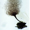 |
Plasmodial
Slime Molds Campbell p.564 Do not confuse this organism with the apicomplexan parasite Plasmodium. These plasmodial slime molds form an ameboid stage that lacks cell walls and feeds on bacteria by engulfing them with pseudopodia- for example, phagocytosis. Plasmodia are found as a slimy wet scum on fallen logs, bark, and other surfaces. |
Barbeyella Metatrichia Sappinia |
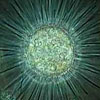 |
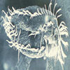
Radiolarians
Campbell p. 563 These heterotrophic protoctists, are distinguished by their long pseudopodia known as axopodia, which are stiffened by microtubules. They feed by engulfing smaller organisms that are trapped on their axopodia. They can range from 30 microns to 2 mm in diameter, and can be found drifting along oceanic currents. |
Acanthocystis Collozoum Thalassicola |
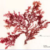 |
Red
Algae (rhodophytes) Campbell p.567 Rhodophytes commonly inhabit the edges of the sea and are cosmopolitan indistribution. In the trophics, particularly, they abound on beaches and rocky shores. The red algae, along with the brown algae are the largest and most complex of the protoctists. Their pigment, phycoerythrin, is what hides the green of chlorophyll. |
Amphiroa Goniotrichum Porphyridium |